In our laboratory we use complex organoids to investigate signaling between epithelial cells and their niche. We use liver as a model organ, where we investigate mechanisms leading to functional decline as a result of aging or disease.
Mammalian organ function and regenerative ability decreases as a result of aging or disease; The liver, which normally possesses an impressive regenerative capacity being able to recover its full size even after 70% resection, is no exception here. We investigate cell communication breakdown in aging and disease of the liver, especially in the context of liver regeneration, and the underlying genetic and metabolic causes of this communication failure.
We utilize complex adult stem cell-derived organoids composed of epithelial cells (hepatocytes, cholangiocytes) and niche cells (such as mesenchyme or endothelium) to understand epithelium-niche cell communication. We combine signaling pathway perturbations with state-of-the art organoid technology, established animal models, tissue staining, live imaging, and single cell sequencing technologies to identify new therapeutic targets in aging and disease.
Prof. Dr. Anna Dowbaj (Principal Investigator)
Ms Isabella Pitzen (Technical Assistant)
Ms Carolin Kraus (PhD Student)
Mr Daniel Veselinovic (PhD Student)
Ms Vi Le (Administrative Assistant)
Human assembloids recapitulate periportal liver tissue in vitro. Yuan L., Dawka S., Kim Y., Liebert A., Rost F., Arnes-Benito R., Baenke F., Götz C., Tsang D., Schuhmann A., Shevchenko A., Rezende de Castro R., Kim S., Sljukic A., Dowbaj A. M., Shevchenko A., Seehofer D., Choi D., Damm G., Stange D. E., Huch M. Nature (2025)
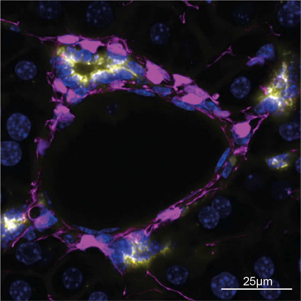
Mouse liver assembloids model periportal architecture and biliary fibrosis. Dowbaj A.M., Sljukic A., Niksic A., Landerer C., Delpierre J., Yang H., Lahree A., Kühn A.C., Beers D., Byrne H.M., Seifert S., Harrington H.A., Zerial M., Huch M. Nature (2025)
Generation of liver mesenchyme and ductal cell organoid co-culture using cell self-aggregation and droplet microfluidics. Dowbaj A.M.*, Kohler T.N.*, Cordero-Espinoza L., Hollfelder F., Huch M. STAR Protocols (2023). *equal contribution
Multisite assessment of reproducibility in high-content cell migration imaging data. Hu J., Serra-Picama X., Bakker G., Van Troys M., Winograd-Katz S., Ege N., Gong X., Didan Y., Grosheva I., Polansky O., Bakkali K., Van Hamme E., van Erp M., Vullings M., Tönisen F., Clucas J., Dowbaj A.M., Sahai E., Ampe C., Geiger B., Friedl P., Bottai M., Strömblad S. Molecular Systems Biology (2023)
Organoids. Zhao Z.*, Chen Z,* Dowbaj A.M.*, Sljukic A.*, Bratlie K.*, Lin L.*, Fong E.L.S., Balachander G.M., Chen Z., Soragni A., Huch M., Zeng Y.A., Wang Q., Yu H. Nature Reviews Methods Primers (2022). *equal contribution
RNF43/ZNRF3 loss predisposes to hepatocellular-carcinoma by impairing liver regeneration and altering the liver lipid metabolic ground-state. Belenguer G., Mastrogiovanni G., Pacini C., Hall Z., Dowbaj A.M., Arnes-Benito R., Sljukic A., Prior N., Kakava S., Bradshaw C.R., Davies S.E., Vacca M., Saeb-Parsy K., Koo B.-K., Huch M. Nature Communications (2022)
Dynamic cell contacts between periportal mesenchyme and ductal epithelium act as a rheostat for liver cell proliferation. Cordero-Espinoza L.*, Dowbaj A.M.*, Kohler T.N., Strauss B., Sarlidou O., Belenguer G., Pacini C., Martins N.P., Dobie R., Wilson-Kanamori J.R. ,Butler R., Prior N. , Serup P., Jug F., Henderson N.C., Hollfelder F., Huch M. Cell Stem Cell (2021). *equal contribution
An optogenetic method for interrogating YAP1 and TAZ nuclear-cytoplasmic shuttling. Dowbaj A.M.*, Jenkins R.P.*, Williamson D., Heddleston J.M., Ciccarelli A., Fallesen T., Hahn K.M., O'Dea R.D, King J.M., Montagner M., Sahai E. Journal of Cell Science (2021). *equal contribution
Quantitative Analysis Reveals that Actin and Src-Family Kinases Regulate Nuclear YAP1 and Its Export. Ege N.*, Dowbaj A.M.*, Jiang M., Howell M., Hooper S., Foster C., Jenkins R.P., Sahai E. Cell Systems (2018). *equal contribution
Full publication list: PubMed, Google Scholar
Prof. Dr. Anna Dowbaj, Chair of Integrated Organoid Systems
E-Mail: anna.dowbaj@tum.de
Admin: Ms Vi Le
E-Mail: vi.le@tum.de; office.dowbaj.cos@bioengineering.tum.de
Phone: 0049-89-289-51703